Detailed imaging without ionizing radiation.
MRI uses strong magnetic fields and radio waves to create detailed images of structures within the body. It can provide valuable information for a wide range of conditions.
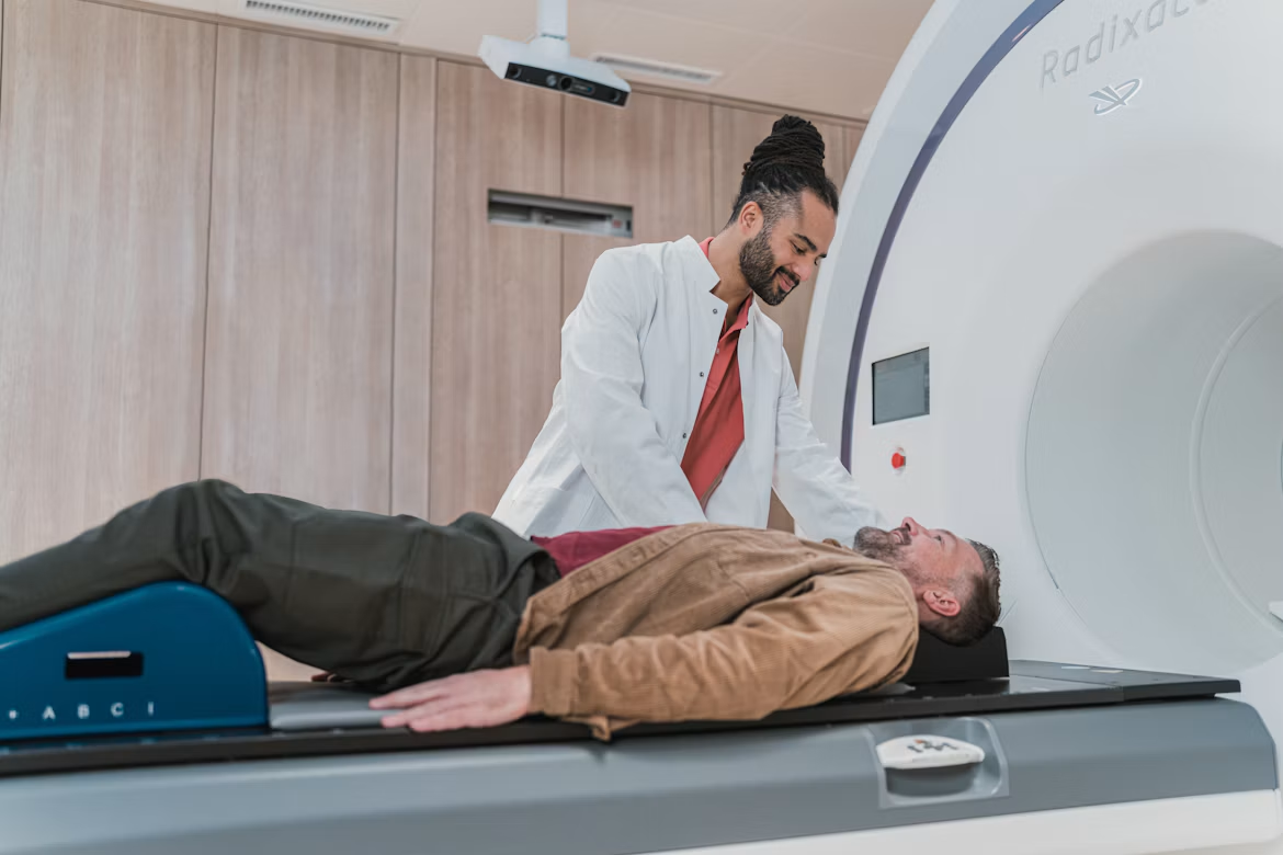
No ionizing radiation.
Understanding MRI
MRI (Magnetic Resonance Imaging) is a non-invasive imaging technique that uses magnetic fields and radio waves to create detailed images of your internal structures. It provides high-resolution views of soft tissues, organs, and the brain without using ionizing radiation.
Because MRI shows soft tissue so clearly, it is often chosen to look at the brain, spinal cord, joints, muscles and internal organs. Your radiologist can compare different image types to understand what is happening.
The scan itself is painless. You lie still on a table that moves into a wide scanner while it records images. The machine is noisy, so we provide ear protection and explain each step before we begin.
What This Imaging Can Help With
Neurological conditions (brain, spine)
Detailed views of the brain and spinal cord to help assess nerve-related symptoms.
Joint and soft tissue injuries
Shows ligaments, tendons, cartilage and muscle that other scans can miss.
Internal organ assessment
Looks closely at organs such as the liver, kidneys and pelvis when more detail is needed.
Cancer detection and monitoring
Helps find abnormalities and track how they respond to treatment over time.
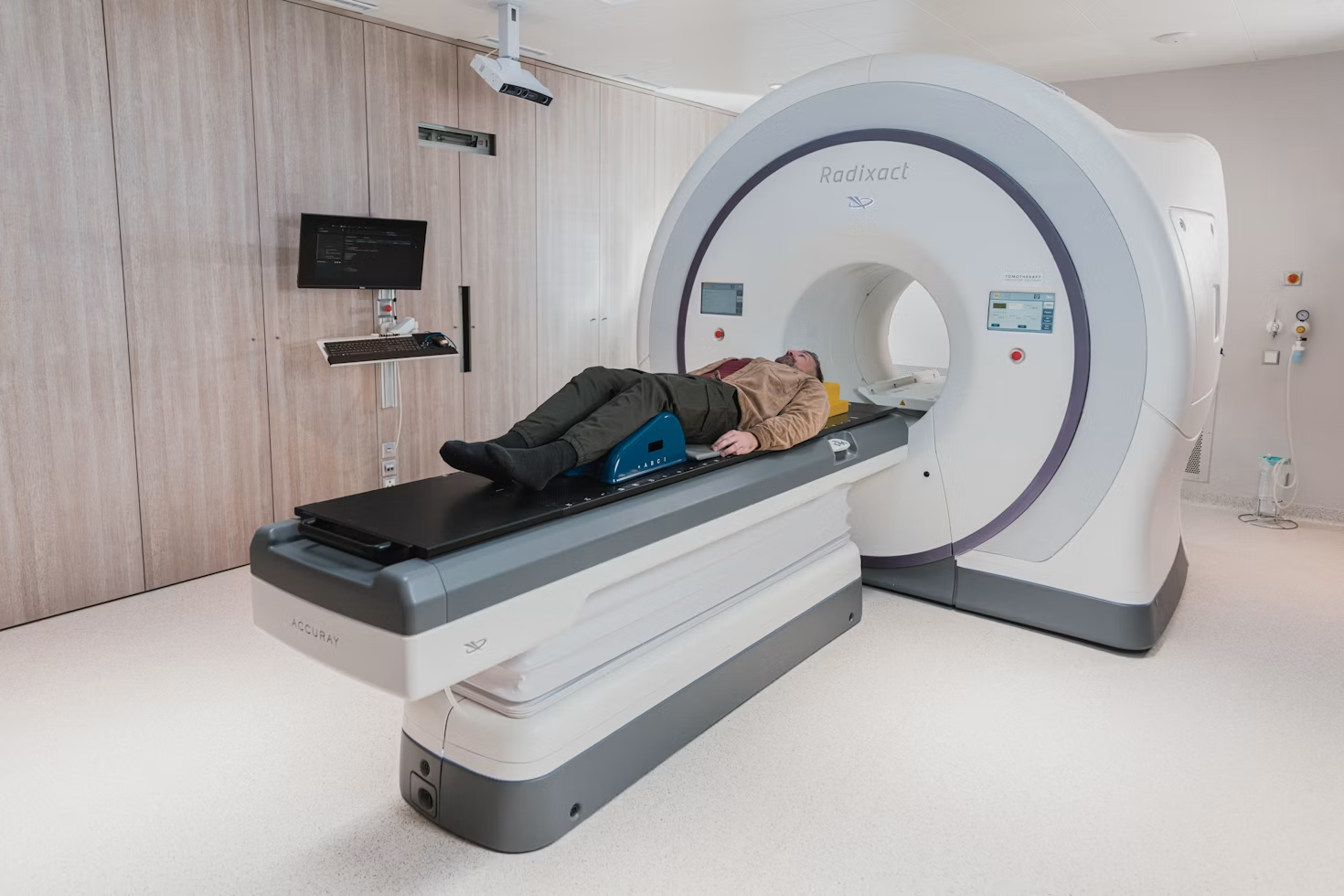
What to Expect During Your MRI
- 01Check-in & preparation(20–30 min)
Confirm your details, complete a safety questionnaire and remove metal items.
- 02Positioning(on the table)
Our team helps you lie comfortably and may place a coil near the scan area.
- 03Imaging process(30–60 min)
The table moves into the scanner. Stay as still as you can and listen for instructions.
- 04Review & analysis(by radiologist)
A radiologist studies your images and prepares a detailed report.
- 05Results(within 1–2 days)
Your report is shared with you or your referring doctor.
Before and After Your Scan
Metal and implants
Tell us about pacemakers, implants, clips or metal fragments before your visit. MRI uses a strong magnet.
Scanner noise
The scanner makes loud knocking sounds. We provide earplugs or headphones.
Staying comfortable
Feel uneasy in enclosed spaces? Let us know and we will help you stay relaxed.
After your scan
You can usually return to normal activities straight away unless advised otherwise.
Book your MRI appointment today.
Our team is here to guide you and answer any questions.
Cross-sectional imaging for accurate diagnosis.
CT scans use X-rays and computer technology to create detailed cross-sectional images of the body. It helps detect a wide range of conditions, from injuries to internal abnormalities.
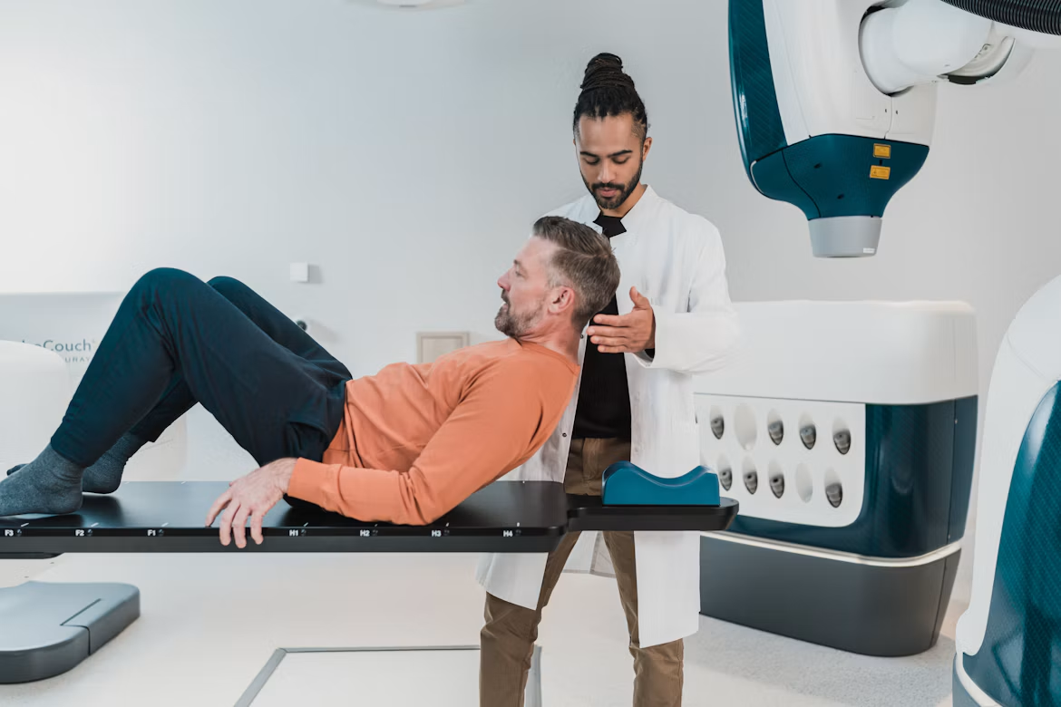
Fast, detailed scans.
Understanding CT Scan
A CT (Computed Tomography) scan combines a series of X-ray images taken from different angles to produce detailed cross-sectional views of bones, blood vessels, and soft tissues. It is fast and widely used for emergency and routine diagnosis.
Because a CT scan takes only minutes, it is often used when a quick answer matters, such as injuries, chest problems or abdominal pain. Images can be viewed as slices or combined into three-dimensional views.
You lie on a table that glides through a short, ring-shaped scanner. The scan is painless and our team stays in contact with you throughout. In some cases a contrast dye is given to help certain structures stand out.
What This Imaging Can Help With
Fractures and bone injuries
Shows breaks, cracks and joint damage quickly and clearly.
Chest and lung conditions
Gives detailed views of the lungs and chest to assess breathing-related concerns.
Internal bleeding and organ assessment
Helps assess organs and detect internal bleeding after injury or illness.
Cancer detection and monitoring
Helps find abnormalities and track how they respond to treatment over time.
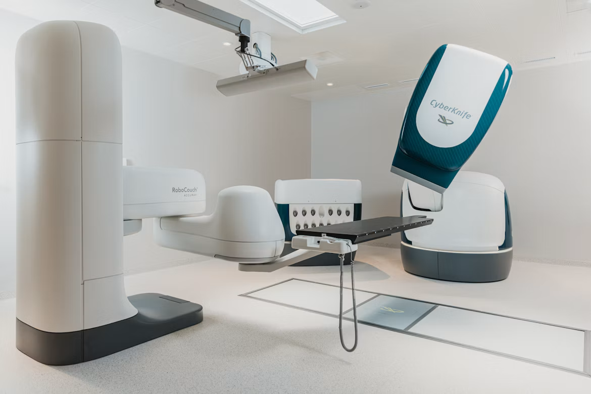
What to Expect During Your CT Scan
- 01Check-in & preparation(10–20 min)
Confirm your details and tell us about allergies or earlier contrast use.
- 02Positioning(on the table)
We help you lie in the right position for the area being scanned.
- 03Imaging process(5–15 min)
The table moves through the scanner. You may be asked to hold your breath briefly.
- 04Review & analysis(by radiologist)
A radiologist studies your images and prepares a detailed report.
- 05Results(within 1–2 days)
Your report is shared with you or your referring doctor.
Before and After Your Scan
Contrast dye
Some scans use contrast. Tell us about allergies, kidney problems or earlier reactions.
Pregnancy
Let us know if you are, or may be, pregnant before your scan.
Eating and drinking
Some scans need fasting. We will tell you if yours does.
After your scan
If you had contrast, drink plenty of fluids unless told otherwise.
Book your CT Scan appointment today.
Our team is here to guide you and answer any questions.
Real-time imaging for better care.
Ultrasound uses high-frequency sound waves to create real-time images of your internal organs, tissues, and blood flow. It is safe, non-invasive, and provides quick results.
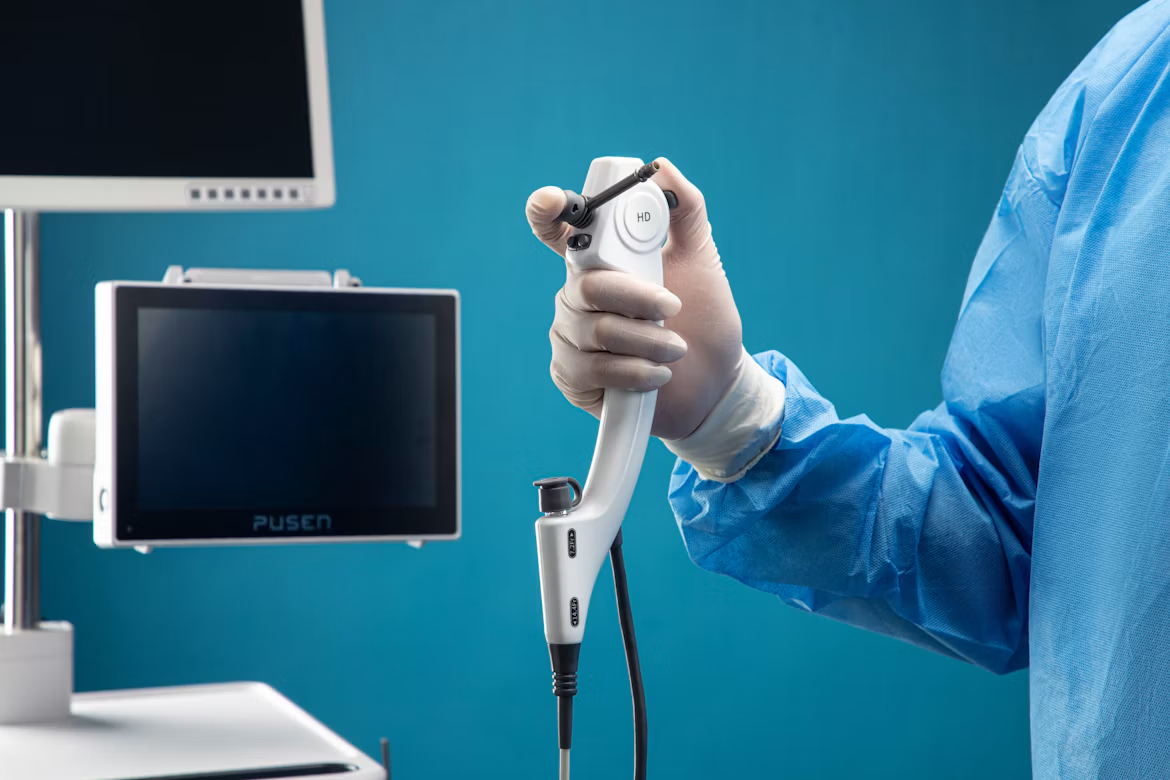
Real-time, radiation-free.
Understanding Ultrasound
Ultrasound (sonography) uses sound waves instead of radiation to produce live images of organs, tissues, and blood flow. It is gentle, comfortable, and commonly used in pregnancy, abdominal, and vascular examinations.
A small handheld device called a transducer is moved gently over the skin with a water-based gel. It sends and receives sound waves, so your sonographer can watch organs, tissues and blood flow as they move.
Ultrasound usually needs little preparation and can often be completed in a single short visit. Our team explains what they are looking at and answers your questions along the way.
What This Imaging Can Help With
Pregnancy monitoring
Follows the growth and wellbeing of your baby without radiation.
Abdominal organ assessment
Examines organs such as the liver, gallbladder, kidneys and spleen.
Blood flow and vascular checks
Shows how blood moves through vessels and can help spot blockages.
Lumps, cysts, and soft tissue findings
Helps tell what a lump or swelling is and decide on next steps.
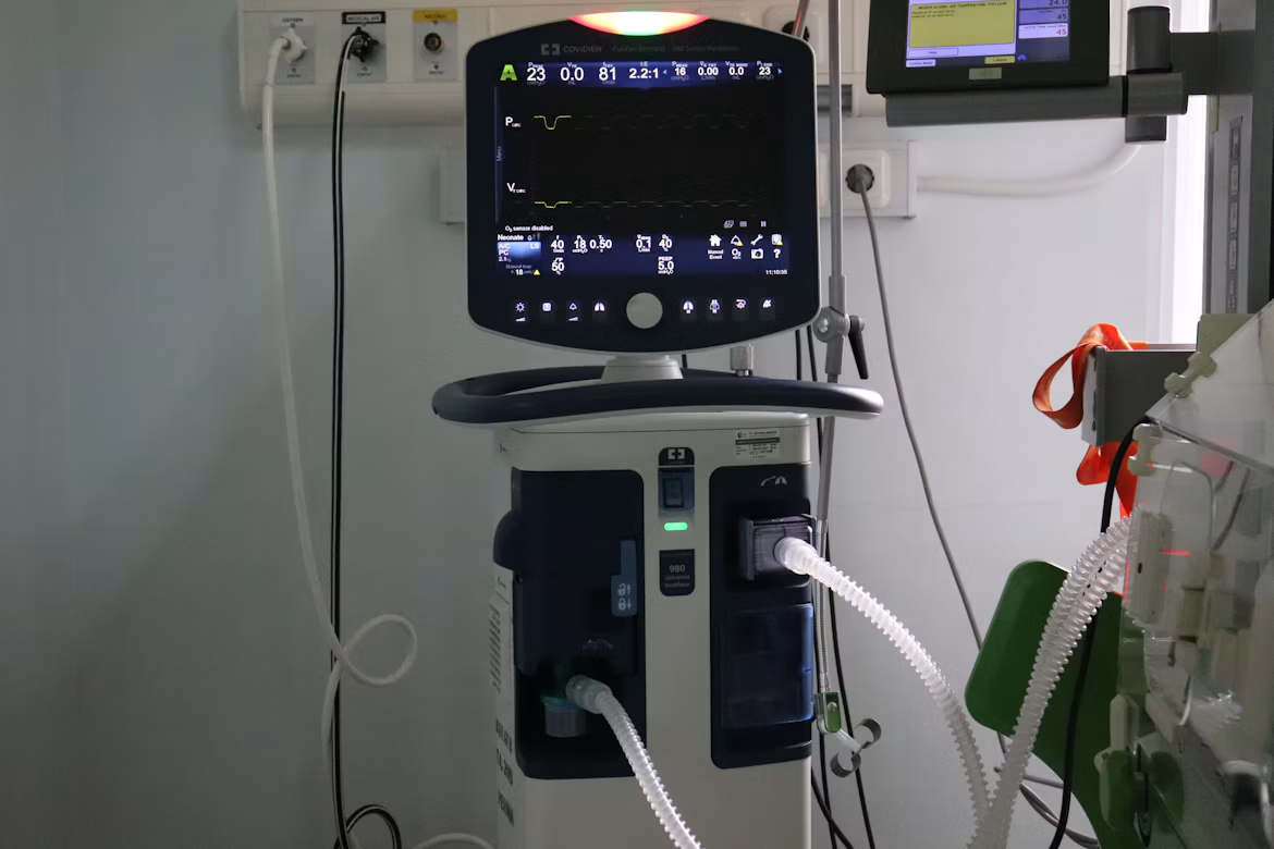
What to Expect During Your Ultrasound
- 01Check-in & preparation(10–15 min)
Confirm your details and follow any bladder or fasting instructions.
- 02Positioning(on the table)
You lie comfortably while a warm gel is applied to the area.
- 03Imaging process(15–30 min)
The sonographer moves the transducer over your skin and captures images.
- 04Review & analysis(by radiologist)
A radiologist studies your images and prepares a detailed report.
- 05Results(same day or next day)
Your report is shared with you or your referring doctor.
Before and After Your Scan
Bladder and fasting
Some scans need a full bladder or fasting. We confirm this when you book.
What to wear
Choose loose, comfortable clothing that is easy to adjust.
Radiation-free
Ultrasound uses no ionizing radiation and is suitable during pregnancy.
After your scan
No recovery time is needed. You can carry on with your day.
Book your Ultrasound appointment today.
Our team is here to guide you and answer any questions.